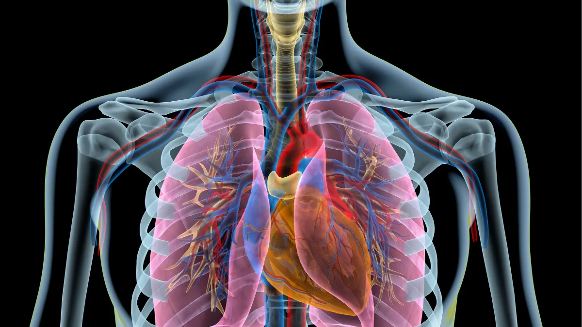
The heart & the lungs: What's the connection?

What’s in a name? #
Consider pulmonary function tests. PFTs are the gold standard for clinicians treating people with breathing problems ranging from asthma to pulmonary fibrosis, and for diagnosing people with dyspnea that doesn’t have a clear etiology. Once a clinician has a better handle on how much gas the lungs are holding, how well that gas is moving in and out, and how well oxygen and carbon dioxide are transferring from the alveoli into the bloodstream, we can start designing personalized interventions to help patients breathe easier.
But lungs obviously cannot exist in a vacuum, literally or figuratively. The work of the lungs can be dramatically affected by any number of organ system dysfunctions. People with issues like chest wall defects or obesity may have mechanical barriers to optimal lung function. Problems in the central nervous system may impact how well the lungs can function, most notably at night. And, of course, trouble in the lungs can affect other organs. There is perhaps no case where this is more true than with the heart, yet PFTs are an often-overlooked tool for the assessment of cardiac conditions.
Welcome to the neighborhood #
When I see those patients who are looking for answers about why they can’t breathe very well, my standard explanation often gets into the difficulties involved in sorting out respiratory conditions from cardiac ones. Many of them start with the same symptoms: Shortness of breath, activity intolerance, weakness, fatigue. Many of them have the same risk factors, including tobacco products or exposure to inhaled pollutants/irritants. In addition to that, there’s a significant amount of collaboration between the two systems; one of my favorite analogies is looking at oxygen delivery as a freight train where hemoglobin “boxcars” get loaded up with oxygen “cargo” and driven to the various tissues by the heart as an “engine.” When the heart’s not pumping well, the oxygen can’t get to where it needs to be, usually causing some degree of subjective dyspnea.
The obvious application of PFTs here would seem to be to try and rule in or out cardiac issues, in order to mover further down the diagnostic path. But again as I tell my patients, the heart and lungs are neighbors in every sense of the word. When you get along with your neighbor, all is well and the neighborhood lives in harmony. When your neighbor starts to throw constant loud parties, it’s a nuisance, but it’s obvious where the nuisance is coming from, and relatively easy to try and address. But what if your neighbor does something subtle, like lets shrubbery slowly grow into your property? It might not be a big deal at first, but if you decide to do some home improvements, you may need to figure out where exactly your property is.
Keep the blood pumping #
That’s why it’s critical to consider the use of PFTs for not only respiratory concerns, but cardiovascular ones. The World Health Organization (WHO) marks cardiovascular disease (CVD) as the #1 leading global cause of death, killing nearly 18 million people around the world every year1. This burden is tremendous, despite years of increasing research and attention focused on combating CVD risk factors, such as high blood pressure and heart failure. One reason why may be the consistent failure to respect the relationship between the heart and the lungs.
Fortunately, research is evolving to highlight this cardiopulmonary connection, and its utility in helping to stay ahead of CVD. One of the more intriguing pieces to come out over the last few years analyzed data from the Jackson Heart Study, a large epidemiological collaboration between the National Institutes of Health, a handful of academic institutions, and the community of Jackson, Mississippi. Designed to identify ways to reduce overall health disparities faced by the Black community and other people of color, the study focuses on reducing the prevalence of CVD at the community level, as well as enhancing care across the continuum. One of the ways the group attempted to do that was through wider use of spirometry.
Spirometry is, of course, a mainstay pulmonary test, frequently used to pick out obstructive lung diseases caused by some CVD risk factors, like tobacco consumption. There’s also a fairly well established relationship between those obstructive diseases and increased burden on the heart, often leading to heart failure (HF) of one flavor or another2. Thus, the result that an obstructive pattern on spirometry was associated with an increased risk for HF. What was a bit of a surprise was that a restrictive pattern was also a distinct risk factor for developing HF, almost as strong as obstruction! The data revealed that after a median follow-up time of 8 years, 8% of those seen with a restrictive pattern were subsequently diagnosed with HF, compared with 10.6% of those with obstruction (and only 3.8% of those with normal spirometry). In addition, the restrictive pattern was associated with higher endothelin levels (a biomarker for CVD risk) and higher pulmonary artery systolic pressure, increasing the risk for pulmonary hypertension34. Again, all of this detected through the use of high-quality spirometry, a tool within reach of any cardiology (or even primary care) practice.
The heartbeat of pulmonary testing #
Additional studies have reinforced the lessons of Jackson. Another wide-ranging epidemiological project, known as the Atherosclerosis Risk in Communities (ARIC) study, was designed to get a better understanding of the risk factors and outcomes of atherosclerosis, another primary risk factor for CVD. In this study, subjects from four cities across the United States (including, coincidentally, Jackson, MS) were followed for several years using various metrics. Baseline spirometry measures were obtained during the subjects’ initial visits, then a follow-up set of readings were taken approximately three years later. The study group found that the quartile of subjects that had the greatest decline in FEV1 between these two measurements were four times as likely to develop heart failure within one year of follow-up, and were also about 25% more likely to experience a stroke4. The news wasn’t much better for those who had rapid declines in their FVC; that group dealt with an elevated risk of heart failure throughout the next 17 or so years of follow-up. These risks held consistent even when accounting for a variety of demographic characteristics, including smoking status, revealing that the lungs can truly provide a look into overall cardiovascular function.
Caring for the overlap #
So far, we’ve looked at cardiac conditions essentially in isolation. But again, many of the same risk factors that lead to heart issues also lead to problems in the lung, and longstanding lung pathologies can certainly have an impact on cardiac function. Thus, we have a high potential for overlap between the two. While debate rages about whether concurrent conditions like asthma and sleep apnea should be considered comorbidities or ‘overlap syndromes,’ any patient-centered approach demands all of these conditions be monitored and managed simultaneously, because it will very often be difficult to predict which process is the primary contributor to mortality. For example, in the case of COPD, many are surprised to realize that mortality is more often attributed to a cardiovascular issue than a pulmonary one2. Thus, it is imperative for primary care providers and cardiologists to maintain a level of comfort with evaluating spirometry data, or (considering the difficulties inherent in care coordination across multiple practices), consider implementing spirometry or pulmonary function testing programs in their own practices. Modern spirometers can fit in the palm of one’s hand, require relatively little maintenance, and require little training time for office clinical staff to become proficient enough in their use to provide high-quality data for clinical decision making. Technology has also advanced to the point where even full pulmonary function testing, once the realm of dedicated laboratories, is available in a compact package, easily compatible with virtually any office that wants the ability to measure every aspect of a patient’s cardiovascular status.
The road ahead #
To be clear, neither spirometry nor any other PFT will ever take the place of traditional cardiac testing, like echocardiography, biomarker evaluation, or the expertise of an experienced clinician. However, much as real-world evidence studies compliment their more rigorous clinical trial counterparts, pulmonary testing can offer even more insight into a person’s overall state. They may be able to provide enough additional information to generate better clinical predictions, and it certainly appears they can guide the management of complex and overlapping disease states. So, as I alluded back at the beginning, perhaps it’s time to revisit the label “pulmonary function test.” Perhaps it’s time to step outside the box a bit and remind clinicians that much as the eyes are considered the windows to the soul, the lungs may be the windows to the thorax, and encourage the use of these procedures to consider problems that originate outside the lungs. There have certainly been rebranding efforts done for less!
Maclay, J. D., McAllister, D. A. & Macnee, W. Cardiovascular risk in chronic obstructive pulmonary disease. Respirology 12, 634-641, doi:10.1111/j.1440-1843.2007.01136.x (2007). ↩︎
Macnee, W., Maclay, J. & McAllister, D. Cardiovascular injury and repair in chronic obstructive pulmonary disease. Proc Am Thorac Soc 5, 824-833, doi:10.1513/pats.200807-071TH (2008). ↩︎ ↩︎
Wang, B. et al. Association of lung function with cardiovascular risk: a cohort study. Respir Res 19, 214, doi:10.1186/s12931-018-0920-y (2018). ↩︎
Morgan, A. D., Zakeri, R. & Quint, J. K. Defining the relationship between COPD and CVD: what are the implications for clinical practice? Ther Adv Respir Dis 12, 1753465817750524, doi:10.1177/1753465817750524 (2018). ↩︎